|
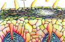
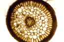
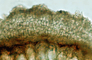
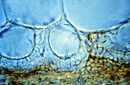
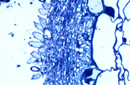
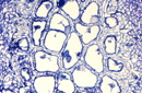
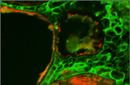
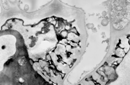
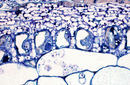
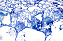
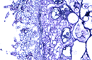
Monotropoid mycorrhizas develop a multi-layered mantle and a paraepidermal Hartig net. Hyphae originating from this or from inner mantle hyphae penetrate into epidermal cells to form fungal pegs. These fungal pegs, one per epidermal cell, are encased by wall material synthesized by the host plant. The wall is laid down unevenly so that each peg has wall projections enveloped by host plasma membrane, a structure similar to that in transfer cells described in many plant tissues. In Monotropa uniflora, the pegs form along the outer tangential wall of epidermal cells, while in Pterospora andromedea, they form along the radial walls. In any cross section of a root, most epidermal cells show the development of these structures. The function of the pegs is not clear, but their structure indicates that they may be involved in nutrient transfer between the fungus and the host.
|